
Medical imaging has revolutionized the way we approach healthcare. It’s like having a magical window that allows us to peek inside the human body without making a single incision.
Among the myriad of imaging techniques available, ultrasound and sonograms stand out as some of the most commonly used diagnostic tools. But what exactly are they, and how do they differ?
The Ultrasound

Definition and Purpose
In the realm of medical diagnostics, refers to the use of high-frequency sound waves to create images of the inside of the body. It’s a non-invasive technique, meaning there’s no need for any surgical procedures to get a good look inside.
The primary purpose? To evaluate and diagnose a variety of conditions, from fetal development during pregnancy to potential problems in organs like the liver, heart, and kidneys.
Fun Fact: The term “ultrasound” refers to sound with a frequency that humans cannot hear. For diagnostic uses, the ultrasound frequency usually ranges between 2 and 18 megahertz (MHz).
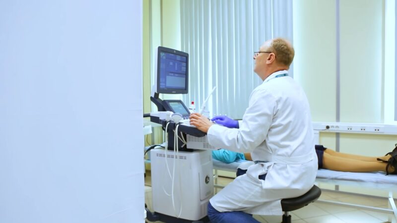
How it Works
At the heart of the ultrasound machine is the transducer – a hand-held device that looks a bit like a wand. The sonographer, the person trained to perform ultrasounds, places this device on the patient’s skin.
As it moves over the body, the transducer emits high-frequency sound waves. These waves travel through soft tissues and fluids but bounce back when they hit denser surfaces.
This echo is what creates the image we see on the screen. It’s a real-time imaging capability, allowing healthcare professionals to see the body’s internal structures in motion.
Pro Tip: The quality of an ultrasound image can vary based on the frequency used. Higher frequencies provide clearer images but don’t penetrate as deeply into the body.
Clinical Applications
Ultrasound isn’t just for expectant mothers wanting a glimpse of their unborn child. It has a wide range of medical applications.
For instance, it can help in evaluating fetal development, detecting problems in organs like the liver, heart, and kidneys, and even assisting in certain types of biopsy procedures. Some common examples include monitoring pregnancies, conducting abdominal scans, and performing vascular studies.
Sonograms

What is a Sonogram?
Now, here’s where things get a tad confusing. The image produced by an ultrasound?
That’s called a sonogram. So, while the terms “ultrasound” and “sonogram” are often tossed around interchangeably, they’re not quite the same thing.
Think of it this way: ultrasound is the process, and the sonogram is the end result – the visual representation of what’s happening inside the body.
Sonogram in Pregnancy
One of the most common scenarios where you’ll hear the term “sonogram” is during pregnancy. These images allow healthcare professionals (and excited parents-to-be) to track the development of the fetus.
They can provide insights into the baby’s health, gender, position, and even sometimes, their mood (yes, babies can be moody in the womb too!). There are different types of prenatal sonograms, including 2D, 3D, and the incredibly detailed 4D scans.
Fun Fact: 4D sonograms are essentially 3D sonograms in motion. They can capture real-time movements, like a baby yawning or stretching.
Diagnostic Sonograms
Beyond pregnancy, sonograms play a crucial role in diagnosing various medical conditions. For instance, they can help determine whether a lump is a potentially cancerous tumor or just a harmless, fluid-filled cyst.
They’re also invaluable in diagnosing issues with soft tissues, muscles, blood vessels, tendons, and joints. Whether it’s investigating conditions like tennis elbow or assessing blood flow in a vessel, sonograms offer a non-invasive way to get the answers needed.
Key Differences

Terminology
It’s easy to get tangled up in the terms “ultrasound” and “sonogram.” While they’re closely related, they’re not identical twins in the world of medical imaging.
- Ultrasound is the actual process, the method of using high-frequency sound waves to peek inside the body.
- Sonogram is the picture or image that results from that process.
It’s like the difference between taking a photograph (the action) and the photograph itself (the result).
Fun Fact: The word “sonogram” comes from the Latin word “sonus,” which means sound, and the Greek word “gramma,” which means something written.
Process vs. Result
To reiterate, ultrasound is all about the action. It’s the technique of sending sound waves into the body, where they bounce off internal structures and return as echoes. These echoes are then converted into the images we see on a screen.
The sonogram, meanwhile, is the visual outcome of this process. It’s the snapshot that doctors, and often patients, look at to understand what’s happening inside.
Pro Tip: If you’re ever unsure about the difference, just remember: You undergo an ultrasound to get a sonogram.
Clinical Usage
In the clinical setting, you might hear doctors use the terms differently based on the context. For instance, a doctor might say, “Let’s order an ultrasound of the liver” when they want to use the imaging technique to look at that organ.
But if they’re discussing the results, they might refer to the “findings on the sonogram.” It’s all about whether they’re talking about the process or the product.
The Role of Both in Healthcare
Complementary Tools
Ultrasound and sonograms are like two sides of the same coin in healthcare. They work hand in hand, with the ultrasound process providing the means to produce the invaluable sonograms that doctors use for diagnosis and treatment planning.
Together, they offer a non-invasive, safe, and efficient way to understand a wide range of medical conditions and guide patient care.
Fun Fact: Ultrasounds aren’t just for humans. Veterinarians use them to examine animals, from household pets to large farm animals.
Advancements and Innovations
The world of medical imaging is always evolving, and both ultrasound technology and sonogram interpretation have seen significant advancements in recent years. Modern machines offer clearer images, 3D and 4D visuals, and even color Doppler to visualize blood flow. These innovations have greatly enhanced diagnostic accuracy and expanded the range of conditions that can be detected and monitored using this technology.
Pro Tip: If you’re due for an ultrasound, it might be worth asking about the latest imaging options available. You might be surprised at the clarity and detail of modern sonograms!
FAQ
What kind of training does a sonographer need?
A sonographer typically requires specialized training, often in the form of a diploma, associate’s degree, or bachelor’s degree in diagnostic medical sonography. Additionally, many countries or states require sonographers to be licensed or certified, which often involves passing an exam.
Are there any risks associated with getting an ultrasound?
Generally considered safe as they do not use ionizing radiation like X-rays. Instead, they use sound waves. However, it’s essential to use them appropriately and only when medically necessary. Prolonged and unnecessary exposure, especially in the hands of an untrained individual, could potentially harm the patient or fetus.
How often can I get an ultrasound during my pregnancy?
The number you’ll need during pregnancy varies based on medical needs. Typically, women have at least two standard ultrasounds: one in the first trimester to confirm the pregnancy and estimate the due date, and one in the second trimester to examine the fetus’s anatomy. Additional ultrasounds might be recommended if there are concerns about the health of the fetus or mother.
Can ultrasounds be used for treatments, not just diagnostics?
Yes, they can also be therapeutic. For instance, they can be used to guide minimally invasive surgical procedures or to break down kidney stones in a process called lithotripsy.
Why do some ultrasounds require a full bladder?
A full bladder can help improve the visibility of certain organs or structures. For instance, during early pregnancy, a full bladder can push the uterus into a better position for imaging. It can also help enhance the image quality by acting as a “window” for the sound waves to pass through, providing clearer images.
Is there a difference in the equipment used for external and internal ultrasounds?
Yes, while the basic principle remains the same, the transducers (or probes) used for internal ultrasounds are designed differently. For example, a transvaginal ultrasound uses a slender transducer that’s inserted into the vagina to get a closer look at the uterus and ovaries. The design and shape of the transducer vary based on the specific internal examination required.
Conclusion
Ultrasounds and sonograms are truly wonders of modern medicine. They provide a window into the body, allowing for early detection, diagnosis, and monitoring of various conditions without the need for invasive procedures.
Recognizing their distinct roles and appreciating their combined contributions can only enhance our understanding and appreciation of these essential diagnostic tools. Here’s to clearer images and a brighter, healthier future for all!